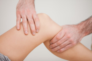
 The knee is a versatile and complicated set of twisting and turning bones, cartilage, ligament and fluid. This joint serves so many purposes – supporting weight through standing, pivoting, running and jumping – that it’s no surprise how many athletes and non-athletes alike are vulnerable to knee injuries and pain. Runners are one group of athletes that are particularly susceptible to injuries from excessive use.
The largest joint in the body, the knee is also one of the most easily injured. It is made up of the lower end of the femur (thighbone), which rotates on the upper end of the tibia (shinbone), and the patella (knee cap), which slides in a groove on the end of the femur. The knee also contains large ligaments, which help control motion by connecting bones and by bracing the joint against abnormal types of motion. Another important structure, the meniscus, is a wedge of soft cartilage between the femur and tibia that serves to cushion the knee and helps it absorb shock during motion.
Common Knee Injuries
Among athletes, knee ligament injuries are common. Sports injuries and even an awkward planting of the foot during a non-athletic activity can result in injuring one of the major ligaments in the knee. The knee is primarily stabilized by pairs of cruciate and collateral ligaments.
The anterior cruciate ligament (ACL) and the posterior cruciate ligament (PCL) cross one another on the inside of the knee. The medial collateral ligament (MCL) is also on the inside of the knee and the lateral collateral ligament (LCL) provides stability from the outside of the knee. Sports related injuries frequently involve the ACL, MCL and PCL.
ACL Injury
For an athlete, an ACL injury is often caused by a quick change in direction, an abrupt change in speed or an awkward landing or pivot. For a football, baseball or soccer athlete, an ACL injury may be caused by the planting or sticking of a cleat in the field of play. A basketball or tennis player may injure the ACL by making a quick change of motion on the court.
Diagnosing ACL injuries may include the need of MRI to determine the severity of the injury. Ligament sprains are frequently described according to a three grades of injury. If the ACL is mildly damaged, but still able to support the knee joint, it’s considered a Grade 1 Sprain. A Grade 2 Sprain describes an ALC where it’s stretched to the point of being loose and a partial tear has been sustained. A complete tear of the ACL is considered a Grade 3 Sprain.
Treatment for ACL injuries is dependent upon the severity of the sprain and the activities required of the patient. Nonsurgical treatment may be recommended for a patient with a partial tear of the ACL, provided the ligament can provide substantial support for the knee. Nonsurgical treatment may include physical therapy and the use of a brace for certain activities.
Surgical treatment for ACL injuries often involve replacing the torn ACL with a graft of tendon taken from elsewhere in the patient’s body. Tendon taken from the patient’s own body is referred to as an autograft. Autograft for ACL replacement is often taken from the patellar tendon, hamstring tendon or quadriceps tendon. Rehabilitation following ACL surgery is crucial. The patient’s commitment to performing exercises and engaging in physical therapy plays an important role in the overall success of the surgical procedure.
MCL Injury
For athletes, MCL injuries are most frequently experienced in contact sports. A collision to the side of the knee during a football, baseball, basketball or soccer match may result in an injured MCL. Such force exerted to the outside of the knee can cause the MCL to stretch to the point of creating a tear. Some MCL tears are isolated and others are part of a complex injury that includes other ligaments, such as the ACL or the meniscus.
Because the MCL has a consistent blood supply, the ligament typically responds well to nonsurgical treatment. Nonsurgical treatment may include resting the knee and wearing a brace to limit side to side movement. Most often, surgery is not required for MCL injuries. When surgery is required, it is typically done through a small incision on the inside of the knee. If the ligament is torn where it attaches to the femur (the thighbone) or tibia (the shinbone), the orthopaedic surgeon will re-attach it to the bone. If the tear is in the middle, the surgeon will sew it together.
PCL Injury
A PCL injury is often the result of strong force, such as a collision, to a knee in the bent position. Twisting or hyperextension of the knee is another way that a PCL injury may occur. Sustaining such an injury can present a patient with difficulty walking and the afflicted kneed may become unstable.
Diagnosing a PCL injury may be done by physical examination combined with an MRI to reveal more about the location and nature of injury. If the PCL is injured solely, without injury to other parts of the knee, nonsurgical treatment, including rest and icing may allow the PCL to heal on its own. A brace may also be used to help immobilize the knee and crutches may be required to limit pressure from weight applied to area. Physical therapy may be recommended as well, to help strengthen leg muscles that support the knee – a proven strategy for assisting the PCL healing process.
If surgery is required, a graft from another part of the body is likely to be used in replacing the torn PCL. Orthopaedic surgeons can perform minimally invasive arthroscopic surgery to rebuild a PCL, allowing for quicker recovery process than traditional surgery. As with other post operative procedures involving joints, the patient’s commitment to physical therapy and dedicated exercises is an important factor in the healing process.
Meniscus Tear
A meniscus tear is often referred to as torn knee cartilage. The meniscus is attached to knee ligaments and acts as a shock absorbing cushion for the knee. Over time, the meniscus is may wear and become more susceptible to tearing. As a sports injury, a meniscus tear is most likely to occur during the action of pivoting, cutting, twisting, being tackled or abrupt deceleration.
In diagnosing a meniscus tear, a physician will perform physical tests that bend, straighten and rotate the knee. MRI may also be required to obtain images of the soft tissues within the knee. Treatment for a meniscus tear is dependent upon the nature and location of the tear, along with other factors, such as patient age and activity demands. Tears occurring within the outside part of the meniscus, where there is rich blood supply, have a strong chance of healing with nonsurgical treatment. Meniscus tears in the areas that lack such a supply of nutrient rich blood are more likely to require surgery. Minimally invasive arthroscopic surgery is typically the procedure performed for meniscus tears that require surgical treatment.
For a severely damaged meniscus, there is the option of meniscal transplant surgery. The primary purpose for meniscal transplant surgery is the replace the meniscus before damage is done to the articular cartilage that protects the knee. When articular cartilage is worn, it can damage the bones moving along the surface, causing pain and leading to a condition of osteoarthritis.
Mensical transplant surgery is performed using arthroscopic surgery, so the healing process is faster than traditional surgery. The transplant surgery uses healthy cartilage tissue from a human donor (a cadaver). Diligent work is done prior to surgery to screen for a donor that is a good match for the procedure. The donor tissue is called an allograft, which is the name for a graft taken from a human donor other than the patient. Patient eligibility for meniscal transplant is dependent on a number of factors, including patient age, overall health and health status of the knee joint being considered.
OCD of the Knee
Osteochondritis dissecans is a joint condition also referred to as OCD, and it is more common to occur in the knee than other joints. It most frequently affects young athletes who have sustained an injury. OCD of the knee is a condition where a piece of cartilage and a layer of the bone beneath it, typically of the femur (the thighbone) comes loose from the end of the bone. X-rays are frequently used to diagnose the condition. Treatment options vary, as the fracture may heal itself with rest, and physical therapy if required.
If nonsurgical treatment proves ineffective, arthroscopic surgery may be used to remove loose fragments or to reattach fragments to the bone. Another surgical procedure, depending on the nature of bone fractures, is to fill the defective area with cartilage containing bundles of collagen fibers known as fibrocartilage. Another option is to use the patient’s own bone marrow to help rebuild the damaged area, encouraging new tissue to grow in the space where a bone fragment is removed.
Articular Cartilage Injury
Articular cartilage is the tissue that covers the ends of bones, allowing bones to move over one another with limited friction. When damaged, either by injury or wear and tear, treatment is typically required, as this cartilage does not usually heal well once compromised. Many of the surgical procedures used to restore articular cartilage are done with minimally invasive arthroscopic surgery.
A variety of surgical options for cartilage restoration exist. One arthroscopic procedure is Microfracture, where multiple holes are created in the joint surface, beneath the cartilage into the subchondral bone. The holes are created using a sharp tool called an awl. This procedure produces a new blood supply reaching the joint surface, delivering new cells that stimulate the growth of new cartilage. Drilling is another arthroscopic option, also aimed at stimulating the growth of healthy cartilage by way of penetrating the subchondral bone. Drilling is done with a small surgical drill or wire. Abrasion arthroplasty is another similar technique, but rather than using drill or wires, high speed burrs are used to simply remove the damaged cartilage and stimulate the healthy subchondral bone.
Autologous Chondrocyte Implantation (ACI) is a two-step procedure used to replace defective cartilage in the knee. ACI is done by extracting healthy cartilage tissue from a non-weightbearing part of the patient’s bone using arthroscopic surgery. The cells from the healthy cartilage are then cultured and increased over a three to five-week period. The second step of the ACI is to conduct an open surgery to implant the newly grown cells into the defective area of cartilage. Prior to implanting the new cells, the cartilage defect is treated with a layer of bone-lining tissue called periosteum, sewn over the area. The newly grown cells are implanted under the periosteal cover.
Two other treatment options for treating Articular cartilage include Osteochondral Autograft Transplantation and Osteochrondral Allograft Transplantation. An Osteochondral Autograft Transplant procedure takes a graft of healthy cartilage tissue from a non-weightbearing area of the patient’s bone, to be utilized in the area of defective cartilage. Because there is a limited amount of area within the patient’s bone to harvest osteochondral autograft, larger transplantation needs are done by way of allograft, where the tissue is taken from a cadaver donor.
Knee injuries may include:
- Knee Instability
- Hearing a loud “pop” noise at time of injury
- Experiencing acute pain inside of knee
- Feeling like the knee is locking or slipping
- Swelling, stiffness or tenderness in or around the joint
Arthritis of the Knee
Arthritis of the knee can be the result of sustained trauma or from disease (osteoarthritis and rheumatoid arthritis). In the case of Osteoarthritis, the deterioration of joint cartilage causes bones to rubs against one another. Arthritis of the knee commonly affects the elderly, overweight individuals and those suffering from pre-existing injury.
Treating Knee Pain
Minimally invasive arthroscopic ACL and PCL reconstruction surgery is a widely accepted treatment for ligament issues. This outpatient technique includes inserting a tiny set of optical fibers and lenses into the affected area. The pictures these lenses send back guides the orthopaedic surgeon to fix or remove the problems causing the pain. Another arthroscopic procedure, meniscus repair, addresses the cartilage problems. Once again, the camera provides images to the surgeon, who then uses sutures to remove, repair or replace the affected cartilage, preventing the tear from widening and causing more pain.
At the Gelb Sports Medicine & Orthopaedic Center, our orthopaedic surgeons are adept at the diagnosis of knee injuries and developing an optimal plan of treatment. Patients benefit from the availability of onsite X-rays and onsite physical therapy.
Total Knee Replacement
The most common condition that leads to total knee replacement surgery is the degenerative joint disease known as osteoarthritis. Typically osteoarthritis affects middle-aged and older adults, though knee trauma and other factors can result in early onset of the disease.
Due to expert training and advancements in medical technology, we most frequently perform total knee replacement surgery with minimally invasive surgical techniques. As a result, patients experience faster recovery time and less pain compared to traditional surgery. The procedure involves removing damaged parts of bone and cartilage and replacing components of the knee with prosthesis. The artificial knee consists of three components, the tibial component (replacing the top of the shin bone), the femoral component (replacing the portion of thighbone) and the patellar component (replacing the bottom surface of the kneecap that rubs against the femur).
Following total knee replacement surgery, the patient will be instructed by physical therapists about the exercises required to assist the healing process.
Partial Knee Replacement
For some patients dealing with the effects of a degenerative disease such as osteoarthritis, a partial need replacement may be an alternative to total knee replacement. In a partial knee replacement procedure, only the damaged part of the knee cartilage is replaced with a prosthesis.
Partial knee replacement surgery may be appropriate for patients with medial, lateral or patellofemoral knee osteoarthritis. In such cases, the area of disease may be localized to the one area of damaged cartilage. Partial knee replacement can be advantageous in that the surgery preserves healthy tissue and bone in the knee, therefore recovery is can be significantly faster.
Expert Treatments Include
- Arthroscopic ACL and PCL reconstruction Surgery
- Arthroscopic meniscus repair using sutures
- Evaluation and treatment of knee conditions including: tendonitis, bursitis, arthritis, meniscus tears, dislocations, instability and sprains
- Evaluation and treatment of fractures of the femur, patella, tibia and fibula
- Total Joint Replacement surgery of the knee
If you’re experiencing pain in the knee or have recently experienced a traumatic knee injury:
Request an appointment online or contact us at 561-558-8898.


